Apr 27 2010
UCLA engineer Aydogan Ozcan has developed a miniature microscope for telemedicine applications based on the Lensless Ultra-wide-field Cell Monitoring Array platform (LUCAS), which is an imaging technology.
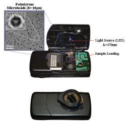 UCLA LUCAS Based Cell Phone prototype
LUCAS uses a digital sensor array for gathering the holographic images of cells or ultrasmall particles, which are generated by an object magnifying lens, along with a light emitting diode (LED) that illuminates the objects. This lens is not utilized for magnifying objects. Images of samples of blood or other fluids can be captured through this technology, even in the countries of the Third World.
The lensless microscope can also be transformed into a Nomarski microscope or a differential interference contrast (DIC) microscope, through the incorporation of in-expensive additional components that cost around $1–2. Compared to traditional microscopes it is lighter in weight and more compact. Since a computer analyzes the images generated and results are immediately made available, the microscope does not require trained mechanics for its operation. Nomarski microscopes are able to provide the sample’s density and places edges and lines in marked contrast to project a 3-D image.
It can be integrated digitally as a constituent of a telemedicine network connecting a hospital or a central lab to many mobile providers of health-care, resulting in filling up the physical infrastructure’s gaps. The cellular networks are already offering the requisite connectivity for these networks, even for the world’s remote regions.
Ozcan hopes that various design elements will render this microscope useful for places with limited resources like some of the African countries. It requires very little training for its operation since the sizeable field of view obviates the requirement to scan the sample, and the sample does not have to be aligned perfectly in the microscope. To operate it, the user has only to fill the blood smear or saliva sample in the chip and place the chip in a slot that is on the microscope’s side. These features render the microscope user-friendly and reliable. It is resistant to problems due to the debris that clog the source of light by virtue of its large aperture.
A self-contained imaging gadget that weighs a mere 46 grams, it can be connected to a computer, PDA, or a smartphone through a USB connection, which also provide power to the microscope and transmit results obtained from uploaded images. It can detect particles and cells along with platelets, white blood cells and red blood cells in the blood sample. The technology will aid in tracking of diseases such as tuberculosis, HIV and malaria in far-off regions at long distances from the healthcare providing facilities. After an earthquake or hurricane, it is also useful for testing the quality of water.
Source: http://www.ucla.edu