In the 21st century, diagnosing illness is essential to prevent untimely deaths and preserve average life expectancy. Image sensors have emerged as a key technology for medical imaging, assisting in the monitoring and clinical diagnosis of disorders. Here, we dive into the fundamentals, applications, and evolution of image sensors in medical imaging.
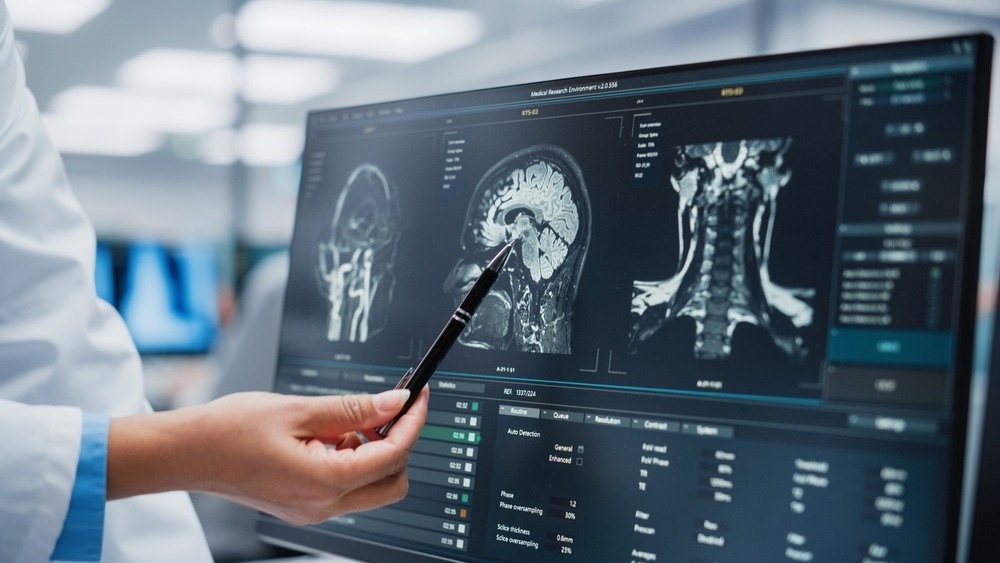
Image Credit: Gorodenkoff/Shutterstock.com
Fundamental Overview of Image Sensors
The conversion of photons into electrons is the fundamental function of an image sensor. The quantum efficiency (QE), which means the efficiency with which an imaging technology converts incoming photons to electrons, is affected by the wavelength of light.
The sensor's sensitivity to light increases with the number of photons converted into electrons, allowing for greater information extraction from the image.
Since a camera may employ filters or a cover glass, the values recorded by the camera could differ from the data provided by the image-sensor supplier.
The market has seen the introduction of two types of image sensors: complementary metal-oxide semiconductor (CMOS)-based image sensors and charge-coupled device (CCD) image sensors.
CCD sensors are commonly found in medical imaging equipment, including endoscopes and X-ray detectors. They are capable of low noise and excellent sensitivity, which makes them ideal for high-quality image capture.
CMOS image sensors have been adopted in most domains of modern technology because of the costly manufacturing process and particular fabrication, as well as the high power usage of CCDs.
The distinction between the sensors is in the way the charge is transported from the pixel to the imaging device or readout. CCD output is analog, but CMOS output is digital. In general, digital cameras are used in life science applications.
What is Medical Imaging?
Medical imaging is used in evaluating several illnesses through imaging. It is critical in validating, analyzing, and documenting the progression of diseases and their response to treatment.
The discovery of X-rays by Wilhelm Roentgen paved the way for advancing medical imaging technologies. Magnetic resonance imaging (MRI), X-ray computed tomography (CT), and other imaging techniques have gradually evolved and been upgraded after years of research.
There are also new imaging technologies coming up, such as infrared, optical, and ultrasonic imaging. Imaging modalities, including X-rays, and ultrasound, may employ diverse kinds of sensors according to their particular imaging properties.
Image sensors have made it possible for medical professionals and researchers to use imaging technologies to obtain accurate and detailed images of human anatomy.
Medical experts use these images to assess and aid in identifying a disease state, which enables the design of a patient-specific treatment strategy.
Image sensors are also employed to observe patients for any potential medical issues that may arise, with uses expanding to advanced surgical methods and routine health screening programs. Healthcare organizations, for example, use screening programs such as enhanced positron emission mammography (PEM) to identify breast cancer.
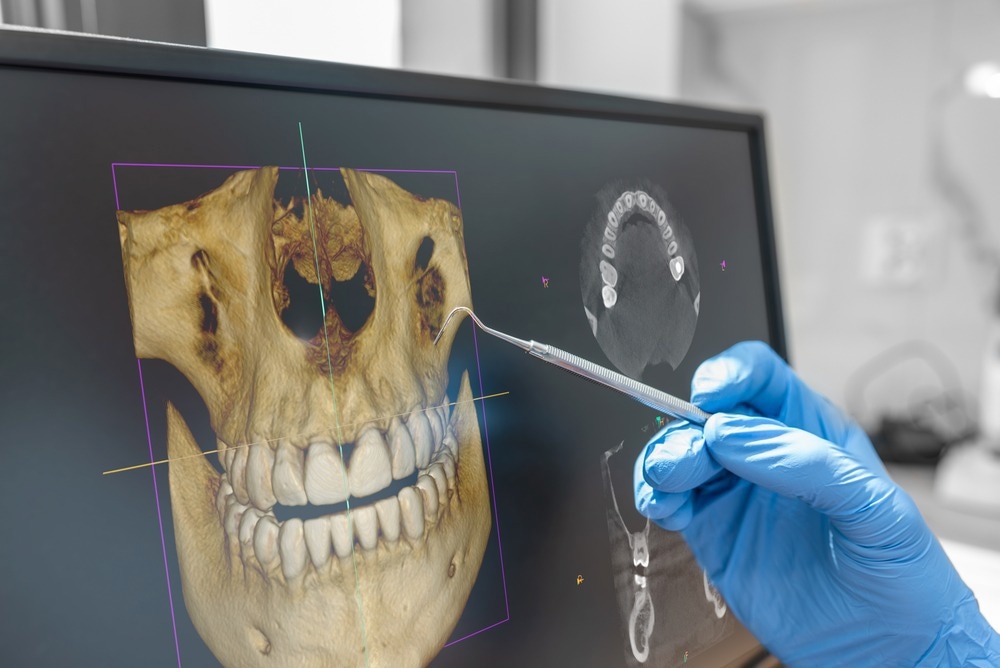
Image Credit: Proxima Studio/Shutterstock.com
Applications of Image Sensors in Medical Imaging
Image sensors have uses in a wide range of life sciences fields, including the ones listed below.
Dental Imaging
Teledyne DALSA has built CMOS-based image sensor technology for dental radiology applications such as panoramic, cone beam CT, and cephalometric imaging.
CMOS image sensors provide major advantages over conventional ones, including radiation hard pixels for a longer image sensor lifetime and package encapsulation method for withstanding adverse conditions, shocks, and vibrations.
X-Ray Image Technology
Teledyne DALSA’s has also created the Xineos X-Ray Image Reconstruction Technology, which bypasses typical limitations by recording clear panoramic images.
The method employs high speed frame based CMOS detectors to capture a 3-dimensional tomographic volume, including therapeutically significant patient anatomy.
Endoscopy and Digital Pathology
In endoscopy, the camera should be small and provide real-time images. The newest CMOS cameras can reproduce even the most delicate color variations and details for surgical imaging systems and endoscopy.
In the field of digital pathology imaging, JAI Apex cameras are optimal for recording cell samples, biopsy tests, and human tissue sections. They can be integrated into microscopes and full-slide scanners.
Another company, Medtronic, created the PillCam SB capsule, which can record two photos per second while traveling through the small bowel for eight hours.
The capsule, measuring 26 mm in length and 11 mm in width, comprises a color video camera along with batteries, a flashlight source, a transmitter, and an antenna.
These video capsules are intended for single use only, have FDA approval, and meet European IEC 60601-1 criteria.
Microscopy
Olympus supplies an array of microscope products to address the demands of life science researchers in a variety of fields, such as drug delivery, cell culture, fluorescence, and neuroscience imaging.
Cameras with CMOS and CCD chips provide high-resolution digital imaging. With super-resolution and excellent three-dimensional images, the microscope can capture the morphological changes and detailed structure of neurons, as well as the deep structure of tissues.
Image Sensors: Current Research
Wiesfield et al. recently created a novel amorphous silicon (Si) image sensor for X-ray medical imaging uses, demonstrating the rapid advancement of medical imaging research.
The devices employ an X-ray phosphor screen attached to a series of a-Si photodiodes for sensing visible light and a-Si thin film transistors (TFTs) for interconnecting the photodiodes to exterior readout circuitry.
They produced images with pixel sizes of 127 mm X 127 mm and imaging areas around 244 mm X 195 mm. These imagers rapidly take pictures and can be connected to a regular PC.
The sensor produced an average QE of almost 70% between 500 - 600 nm, with a light-sensitive area of 57% and optimal QE for x-ray phosphor emission in the green region of the spectrum.
Another report published in Review of Scientific Instruments, stated the development of a simple, and reliable method of using charge coupled device as an X-ray fluorescence (XRF) detector. The key to this technology is that X-ray photons produce charge in the charge coupled device-based chip, resulting in a rapid readability.
The results demonstrated that full field XRF imaging possesses a high spatial resolution. Full field XRF imaging is thus possible when a normal CCD camera is combined with a pinhole collimator.
Future Outlook
Medical science has been transitioning from conventional imaging practices to computer-aided medicine due to the constantly developing state of smart technologies like image sensors.
Such devices have produced innovative and accessible strategies as well as state-of-the-art techniques for paradigm shifts in diagnostic and therapeutic approaches.
Medical imaging sensor development will progress in two directions in the future. The first path is "intelligent". An intelligent sensor is a mix of sensitive components, namely communication circuits, micro-processors, and intelligent software systems.
The second course of action is miniaturization. Sensor technology will move from micro-sensors to nano-sensors as micro-electronic processing technology, particularly nano-processing technology, advances.
References and Further Reading
Microscopes | Olympus LS. [Online] Available at: https://www.olympus-lifescience.com/en/microscopes/
C. Connolly, (2005) Imaging developments benefit medical applications, Sens. Rev., vol. 25, no. 4, pp. 246–248, doi: 10.1108/02602280510620079/FULL/XML.
K. Di Feng, Z. Wang, and Y. Yang, (2020) Development of medical imaging sensors, Int. J. Distrib. Sens. Networks, vol. 16, no. 1, doi: 10.1177/1550147720903607/ASSET/IMAGES/LARGE/10.1177_1550147720903607-FIG20.JPEG.
Y. Shi and Z. Liu, (2023) Evolution from Medical Imaging to Visualized Medicine Adv. Exp. Med. Biol., vol. 1199, pp. 1–13, doi: 10.1007/978-981-32-9902-3_1/COVER.
W. Zhao and K. Sakurai, (2017) CCD camera as feasible large-area-size X-ray detector for X-ray fluorescence spectroscopy and imaging,” Rev. Sci. Instrum., vol. 88, no. 6, doi: 10.1063/1.4985149/361288.
Operating principle and features of CMOS sensors | Baumer India. [Online] Available at: https://www.baumer.com/in/en/service-support/function-principle/operating-principle-and-features-of-cmos-sensors/a/EMVA1288
Applications of CMOS in Medical Imaging and Life Sciences. [Online] Available at: https://www.kolabtree.com/blog/applications-of-cmos-in-medical-imaging-and-life-sciences/
Types of Medical Scanners in diagnostic imaging|Open Medscience. [Online] Available at: https://openmedscience.com/what-is-medical-imaging/
GHO DEVICES08 Total density per million population: Magnetic Resonance Imaging MAP TEMPLATE. [Online] Available at: https://www.who.int/activities/strengthening-medical-imaging (accessed Jan. 07, 2024).
R. L. Weisfield, M. A. Hartney, R. A. Street, and R. B. Apte, “New amorphous-silicon image sensor for x-ray diagnostic medical imaging applications,” https://doi.org/10.1117/12.317044, vol. 3336, pp. 444–452, Jul. 1998, doi: 10.1117/12.317044.
Home | Teledyne DALSA. [Online] Available at: https://www.teledynedalsa.com/en/home/
JAI | JAI cameras for supreme color imaging in medical and life…. [Online] Available at: https://www.jai.com/markets-applications/medical-and-life-sciences
PillCamTM SB 3 Capsule Endoscopy System | Medtronic. [Online] Available at: https://www.medtronic.com/covidien/en-us/products/capsule-endoscopy/pillcam-sb3-system.html
Disclaimer: The views expressed here are those of the author expressed in their private capacity and do not necessarily represent the views of AZoM.com Limited T/A AZoNetwork the owner and operator of this website. This disclaimer forms part of the Terms and conditions of use of this website.