Current endoscopic techniques allow doctors to carry out surgeries without performing any extreme incision, however, certain surgeries require tools with specific 3-D optics. An image sensor that has the capacity to transmit accurate 3-D images from within the human body has been developed by researchers.
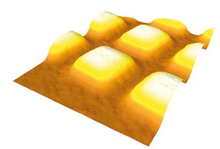
 Specially designed microlenses will one day help to transmit 3-D images from inside the human body. Their function is to precisely focus the light rays on the sensor.
Through the nasal cavity of the patient, the surgeon carefully directs the endoscope to the operation area. In this very delicate technique, the surgeon has to carry out detailed preparation before starting the actual intervention. The camera, incorporated in the slender endoscope tube, allows the surgeon to view every detail in clear 3-D resolution. The stereoscopic vision furnished by a 3-D endoscope makes the operation of neurosurgeons and other experts significantly simpler. The 3-D endoscope can steer securely through the tissue without creating collateral damage and helps complete the work faster.
Scientists at the Fraunhofer Institute for Microelectronic Circuits and Systems IMS in Duisburg and the design partners in the EU scheme “Minisurg” have developed the new image sensors. The currently available CCD sensors offer only low-resolution images whereas the new CMOS image sensors, created by the researchers, are similar to the sensors commonly included in single-lens-reflex (SLR) cameras and are suitable for medical applications.
In order to make the sensors suitable for medical applications, the researchers created unique microlenses that are incorporated in the CMOS sensors. In the CMOS sensors, a cylindrical microlens is fixed before every two perpendicular lines of sensors in the pixel arrangement. A patterned lens captures the light focused on the microlenses, which directs it on the pixels. The special feature of this configuration is that the two light rays are caught by the lenses. Accordingly, the CMOS sensor gets two groups of image information that are handled separately in a manner similar to how the brain treats the images coming from the right and left eye. The surgeon can either view the 3-D images on the screen directly or by using polarized glasses.
Researchers had to satisfy a number of challenges relating to the creation of a miniature camera by producing a chip that can fit into the tube that does not exceed over 7.5 mm in diameter. Along with the bunch of optical fibers that operates as the light source, the diameter of the endoscope is 10 mm.
Source: http://www.fraunhofer.de/en.html