University of Washington researchers, along with their Stanford University colleague, have designed biosensors that can observe the manner in which a message is conveyed to a bacterium to result in dividing it into two structurally and functionally different cells. These biosensors are capable of measuring biochemical fluctuations within a single bacteria cell whose size is smaller than a plant or an animal cell.
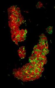 UW This image is the first visualization of a second messenger through a biosensor in bacteria, which are much smaller than animal or plant cells.
These scientists analyzed the cell division existing in disease bacteria’s species, which is able to fend off treatment and uses these defenses, known as Pseudomonas aeruginosa, to establish a stronghold. This rod-shaped pathogen is the cause for the life-reducing, chronic lung infections in patients suffering from cancer-associated suppressed immune systems, cystic fibrosis, and burns. These researchers also studied cell-division existing in a safe lake and stream dwelling bacteria, known as Caulobacter crescentus. The Science journal’s June 4 issue will be publish the researcher’s findings.
For monitoring the c-di-GMP’s concentration inside single living bacteria cells, a biosensor was developed by the scientists. This biosensor is built up using the fluorescence resonance energy transfer that is genetically encoded.
C-di-GMP is able to control several biological functions in the cell by connecting up with a dissimilar array of receptors. These include proteins for building and driving hair-like, waving, structures for moving cell. These also incorporate proteins, riboswitches – RNA molecules, and transcription factors which are able to change the activity of the gene.
Due to the C-di-GMP’s ability to control various different cell functions, the researchers were lead to believe it was quite possible that it is able to manage its regulatory workload by being present at the right time, at the right amount, and at the right place in the cell cycle.
The National Institutes of Health’s National Institute of Allergy and Infectious Diseases, the Swiss National Foundation, the National Science Foundation’s graduate research fellowship, the Novartis Foundation, and the Cystic Fibrosis Foundation provided grants to support the research.