The art of tattooing may well enter the field of medical diagnosis. Researchers in Germany have designed permanent dermal sensors that can be applied as artistic tattoos. As reported in the journal Angewandte Chemie, instead of tattoo ink, a colorimetric analytic formulation was injected into the skin. The pigmented skin areas changed their color when blood pH or other health indicators differed.
 © Wiley-VCH
A tattooist puts ink directly in the dermis, an approximately one-millimeter-thick layer of tissue where blood vessels, nerves, and hair follicles are found. The tattoo needle punctures the epidermis, the topmost layer of skin, and discharges the pigments into the dermis below, where the pigments form a permanent stain the skin.
Using tattoos for diagnostic instead of cosmetic purposes is a new theory. Researcher Ali K. Yetisen, who is employed at the Technical University of Munich, Germany, and his coworkers thought the method could be useful to place sensor formulations at areas in the body where they can record variations in metabolic substances directly, without any time delay or spatial distance, and possibly for an extended period of time.
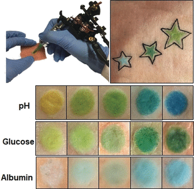
The scientists then found and adapted three colorimetric chemical sensors that create a color change in reaction to biomarkers. The first sensor was quite a simple pH indicator containing the dyes bromothymol blue, methyl red, and phenolphthalein. If injected into a model skin patch such as a piece of pig skin, the resulting tattoo changed from yellow to blue if the pH was modified from five to nine.
The other two sensors investigated the levels of albumin and glucose. Albumin is a carrier and conveys protein in the blood. High glucose levels in the body may specify diabetic dysfunction, while dropping albumin levels can specify kidney or liver failure. The glucose sensor consists of the enzymatic reactions of glucose oxidase and peroxidase, which subject to the glucose concentration resulted in a structural variation of an organic pigment, and a yellow to dark green color change. The albumin sensor was based on a yellow dye that, upon exposure to the albumin protein, changed to green.
The researchers then applied many sensor tattoos onto patches of pig skin. When they altered the pH or the albumin or glucose concentrations, the colors of the decorated areas varied accordingly. They quantified these observable effects by assessing the colors with a basic smartphone camera and an app.
The researchers state that such sensor tattoos could enable permanent tracking of patients using a basic, low-cost method. With the creation of appropriate colorimetric sensors, the method could also be used for recording pathogen and electrolyte concentrations or the dehydration level of a patient. More studies will investigate whether tattoo artwork can be used in a diagnostic setting.