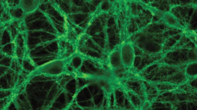
Confocal image of cultured neurons expressing OxLight1. (Image Credit: University of Zurich).
Now, researchers at the University of Zurich have created a fluorescent orexin biosensor to observe this molecule "live" in the living mouse brain.
Billions of neural cells in the brain act in unison to coordinate both routine and higher functions of an organism.
Neural cells use a distinctive language to communicate with each other: molecules called neurotransmitters or neuropeptides. Orexin is an example of one such signaling molecule system.
Typically, it controls motivation, wakefulness, arousal and appetite. Deficiencies in the sensing or release of orexin neuropeptides result in, both in animals and in humans, a disease known as narcolepsy.
Affected individuals have to deal with overwhelming daytime sleepiness and frequently display cataplexic states, in which they stay conscious but are not able to regulate body movement, resulting in a kind of paralysis.
Shedding Light on the Internal Workings of the Mouse Brain
Tommaso Patriarchi from the Institute of Pharmacology and Toxicology at the University of Zurich (UZH) and his team have currently designed a genetically encoded biosensor whose fluorescent properties allow them to examine, in high-resolution, orexin action and release processes "live" in the brain of living mice.
The direct link between this particular neuropeptide system and its dramatic alteration in human brain functions in narcolepsy, led us to study orexin in more detail.
Tommaso Patriarchi, Institute of Pharmacology and Toxicology, University of Zurich
The new orexin biosensor named "OxLight1" is established on a specially engineered green fluorescent protein combined into the human orexin receptor.
"Marking the receptor with a fluorescent protein makes it visible under the microscope. When the neuropeptide binds to the receptor, it makes it light up", Patriarchi adds.
OxLight1, therefore, essentially provides a real-time view of orexin release in living animals such as a mouse.
Illuminating Previously Invisible Aspects of Healthy Brain Function
To understand how neuropeptides systems like orexin act to maintain a healthy brain function, we need to be able to first observe the messages carried by these neuropeptides and then learn to interpret them.
Tommaso Patriarchi, Institute of Pharmacology and Toxicology, University of Zurich
Until now, this has been virtually impossible because of the lack of tools available to offer a readout with high temporal and spatial resolution. The scientists, thus, employed their new biosensor to examine the relationship between neuronal activity and neuropeptide release in living animals, one of the most demanding and long-pursued questions in neurophysiology that has remained intangible thus far.
They demonstrated that the level of orexin release correlates with both duration and frequency of neuronal activation.
"The exquisite sensitivity and speed of OxLight1 allowed us to track endogenous orexin release associated with natural behaviors such as spontaneous running or acute stress", says Patriarchi.
Thus, they were able to show in the living brain that orexin signals can take place in the form of comparatively short-lived or "phasic" spurts of release.
Investigating the Neural Disease Mechanisms of Narcolepsy
The researchers then examined orexins dynamics in sleep/wake transitions. By integrating photometry imaging of orexin dynamics and neuronal activity recordings to rate the sleep status of the animals, the team noticed, for the first time, that a swift drop in orexin levels happens during REM sleep of the mice.
Further work with colleagues from the Istituto Italiano di Tecnologia in Italy, experts in two-photon microscopy, exposed another so far unfamiliar process: spatially localized orexin fluctuations taking place in the somatosensory cortex when awakening from anesthesia. This latter joint research was carried out within the framework of the recently awarded European project DEEPER.
After deciphering orexin release and neuronal activity in the healthy brain, we are now using OxLight1 to investigate the mechanisms of brain diseases like narcolepsy and addiction.
Tommaso Patriarchi, Institute of Pharmacology and Toxicology, University of Zurich
This study is the first result of a project for which Tommaso Patriarchi was given an ERC Starting Grant in 2020. The biosensors that his team has engineered are currently being used to examine brain function in laboratories worldwide. By continuing to increase their neuro-technological toolbox, the scientists also hope to create advanced screening assays for drug development.
Journal Reference:
Duffet, L., et al. (2022) A genetically encoded sensor for in vivo imaging of orexin neuropeptides. Nature Methods. Nature Methods. doi.org/10.1038/s41592-021-01390-2.