A new study from researchers at the University of British Columbia (UBC) introduces a living biosensor built from gut bacteria that dims its fluorescent glow when it detects signs of disease, offering a promising new way to monitor gastrointestinal health non-invasively.
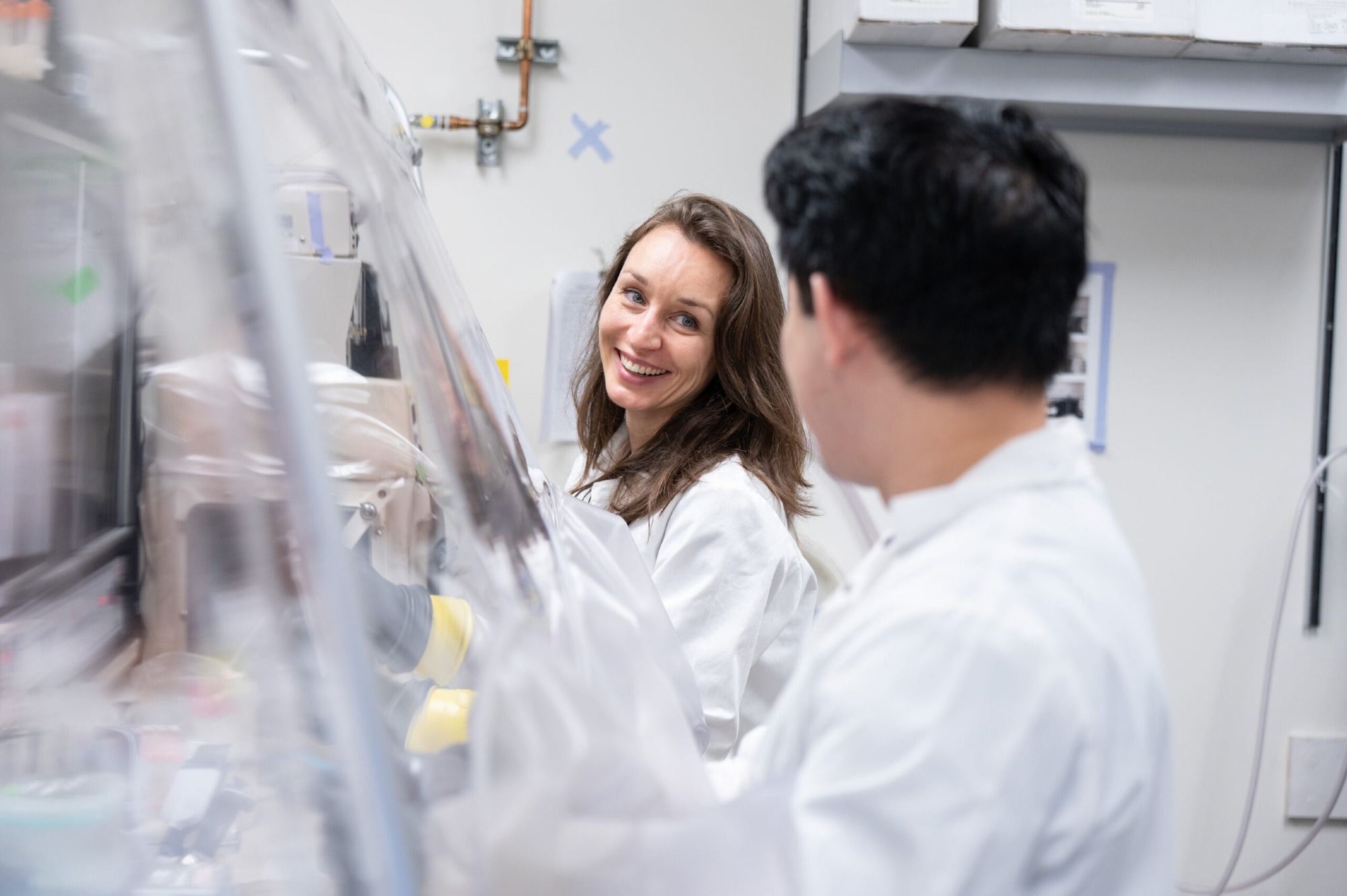 Dr. Carolina Tropini, senior author of the study and assistant professor in the University of British Columbia's Department of Microbiology and Immunology, and the School of Biomedical Engineering. Image Credit: University of British Columbia
Published in Cell, the research presents a tool that could dramatically improve how we track gut conditions by using bacteria already present in the digestive system.
Our biosensors could improve the ability to predict how diseases in the gut progress, identifying early changes that could aid preventative interventions.
Juan Camilo Burckhardt, Study Co-First Author and Doctoral Candidate, Department of Microbiology and Immunology (MBIM), University of British Columbia
Currently, diagnosing gut-related diseases often requires invasive procedures that provide only a single snapshot in time. In contrast, the biosensor developed by UBC researchers is designed to continuously monitor gut conditions (by analyzing stool samples) without disrupting the natural gut environment.
Utilizing ‘Good’ Gut Bacteria
Beneficial bacteria that naturally reside in the intestine and support gut health are highly sensitive to local conditions and have evolved to thrive long-term in these environments. Building biosensors in these bacteria, therefore allows researchers to continuously monitor the gut environment without disturbing it.
Dr. Giselle McCallum, Study First Co-author and Doctoral Student, University of British Columbia
By identifying genes in B. theta that respond to common disruptions, such as those seen in celiac disease and inflammatory bowel disease, the team created a tool that reacts to subtle environmental changes in the gut.
One key marker they tracked is osmotic stress, a condition where poor nutrient absorption leads to a buildup of undigested molecules. These molecules pull excess water into the intestines, often triggering diarrhea and inflammation, which can aggravate underlying conditions.
Understanding these gut changes is essential for advancing our diagnostic and treatment strategies for gut health. For that, we need highly sensitive measurements as those changes occur, including before symptoms appear.
Dr. Carolina Tropini, Study Senior Author and Assistant Professor, School of Biomedical Engineering, University of British Columbia
Linking Glowing Proteins
Traditional biosensors typically rely on bacteria glowing in response to stress, but B. theta’s glow wasn’t bright enough to work this way. Instead, the team flipped the model: they engineered the bacteria to glow under healthy conditions, and dim when stressed. That way, a decrease in brightness acts as a clear signal that something is off.
Testing this approach in mice, the researchers tracked the glow in bacterial cells collected from stool samples.
“We found that the biosensor accurately reported osmotic stress in the gut, even picking up subtle changes that didn’t cause clinical symptoms like diarrhea. It remained stable and responsive for weeks, which means it could track the gut environment long-term and potentially detect illness before symptoms develop,” said Burckhardt.
The research team is now working on adapting the biosensor to monitor other gut-related factors, including oxygen levels, temperature, and pH.
“While early applications will likely focus on monitoring gastrointestinal diseases, the long-term goal is a personalized approach where people can track aspects of their gut health over time and identify early warning signs of imbalance or dysfunction,” said Dr. Tropini.
Looking ahead, the team envisions biosensors that do even more, like releasing medications only when specific disease-related changes are detected.
Journal Reference:
Tropini, C., et al. (2026) A Bacteroides synthetic biology toolkit to build an in vivo malabsorption biosensor. Cell. DOI: 10.1016/j.cell.2025.12.052. https://www.cell.com/cell/abstract/S0092-8674(25)01501-6